
Printable Skull Anatomy Coloring Pages
These 24 printable skull anatomy pages provide accurate diagrams for biology students, teachers, and adult colorists. You will find detailed illustrations of cranial bones, suture lines, and internal views to help you study the human head.
Human Skull Anatomy Coloring Pages
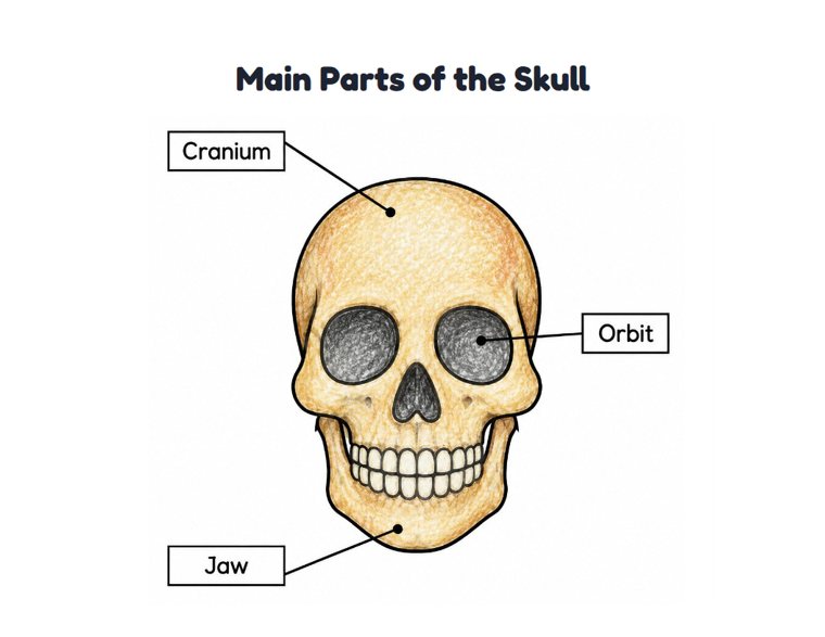
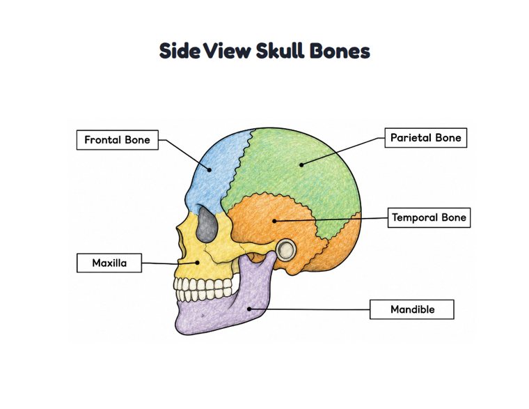
Explore standard anterior and lateral perspectives showing the major cranial bones and distinct suture lines.


Labeled Skull Diagrams and Study Worksheets
Use these numbered worksheets and labeled illustrations to test your knowledge of cranial terminology.


Inferior Base and Internal Cranial Views
Study the complex structures of the cranial fossae and the inferior base of the cranium.


Facial Skeleton and Jaw Bone Outlines
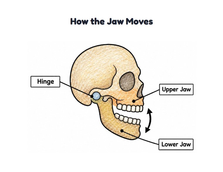
Focus on the lower jaw and the intricate bones that make up the human facial structure.


More pages to explore
Related coloring pages on Color Confetti.


All 14 more: Skull (40), Anatomy (29), Science (28), Tooth (31), Skeleton (17), Brain (29), Human (33), Human Heart (24), Heart Anatomy (25), Circulatory System (21), Nervous System (23), Digestive System (21), Cell (16) and Doctor (26).
Ready to print for home, preschool and classroom.
PDF paper size:
Using these pages in your nonprofit, library, hospital or at an event? We'd love to hear from you
Skull Anatomy Worksheets
5 printable skull anatomy worksheets with answer keys, from tracing first words in Pre-K to labeling and writing in grades 3–4. Every PDF has the worksheet, the answer key and a colored example. You can change the title and the words before printing.
Download all 5 worksheets as one PDF with answer keysMajor Bones of the Skull in 2 levels
More skull anatomy worksheets
Understanding Cranial and Facial Bones
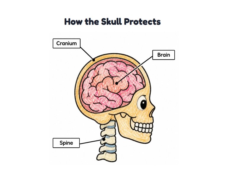
The human skull is a complex structure made up of 22 individual bones. When studying these anatomy coloring pages, it helps to divide them into two main groups. The neurocranium forms the protective vault around the brain, including the frontal, parietal, temporal, and occipital bones.
The viscerocranium creates the framework of the face, housing the zygomatic bones and the maxilla. Coloring these distinct areas helps students memorize their spatial relationships. Using a consistent color key across different views reinforces learning.
Navigating Sutures and Internal Structures
One of the most challenging aspects of studying the head is understanding how the bones connect. The fibrous joints, known as cranial sutures, are clearly outlined in our labeled study worksheets. Tracing the coronal, sagittal, and lambdoid sutures with a fine-tip pen can help you visualize these boundaries.
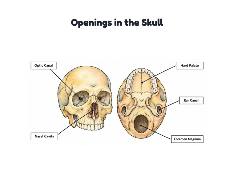
For advanced students, the internal cranial views reveal the intricate pathways for nerves and blood vessels. You can highlight the anterior, middle, and posterior cranial fossae using subtle shading techniques to understand the depth and contours of the skull base.
Educational Uses for Teachers and Students
These printables are designed for academic use in science classrooms and home study sessions. Biology teachers can use the numbered diagrams as quiz materials or homework assignments. Medical and nursing students often find that physically coloring the structures improves retention of complex medical terminology.
- Assign the exploded skull diagrams to show how individual bones articulate.
- Use the mandible and teeth illustrations to discuss dental anatomy.
- Print the cross-section views to help students understand the relationship between the skull and the cervical spine.
Coloring Techniques for Anatomical Diagrams
When completing these diagrams, precision is more important than artistic flair. Adult colorists and students should choose colored pencils with firm leads that can stay sharp. This allows you to stay within the narrow lines of the foramina and small facial bones.
Highlighters or light markers work well for the larger flat bones, ensuring that any printed text or numbering remains visible underneath the color as you study the human skeletal system.